


Abdominal Muscles
Abdominal muscles, from an aesthetic perspective, are often regarded as one of the most desirable muscle groups to develop. For many, the term "abs" typically refers to the "six-pack," which is just a fraction of the abdominal region. The functionality of the abdominal muscles goes far beyond their aesthetic appeal, playing a significant role in spinal stability which is crucial for both everyday activities and athletic performance. The abdominal muscle group comprises four main muscles: the rectus abdominis, external obliques, internal obliques, and transversus abdominis. When these muscles contract, they enable trunk flexion and rotation and help prevent anterior pelvic tilt. These muscles also play a pivotal role in the movement and stabilization of the lumbar spine and control the movements between the pelvis and spine. Weak abdominal muscles can lead to anterior pelvic tilt, increased lumbar lordosis, and a higher risk of lower back pain.
Anterolateral Group
The anterolateral group forms the anterolateral wall of the abdominal cavity, including the external oblique, internal oblique, transversus abdominis, and rectus abdominis muscles.
External Oblique
The external oblique muscle, located on the superficial layer of the anterolateral abdominal wall, is a broad, flat muscle. It originates from the outer surfaces of the lower eight ribs, interdigitating with the serratus anterior and latissimus dorsi muscles, with its fibers running diagonally downward and medially. The lower posterior fibers terminate at the anterior part of the iliac crest, while the remaining fibers transition into an aponeurosis that covers the rectus abdominis anteriorly, contributing to the anterior layer of the rectus sheath and terminating at the linea alba. The inguinal ligament, formed by the thickened lower edge of the external oblique aponeurosis, extends between the anterior superior iliac spine and the pubic tubercle. The lacunar ligament is a small bundle of fibers extending from the inguinal ligament, turning posteriorly and downward to the pecten pubis, also known as the pectineal ligament or Cooper's ligament. Both the inguinal and pectineal ligaments are vital structures used to reinforce the inguinal canal wall during hernia repair surgeries. Above the pubic tubercle, the aponeurosis of the external oblique forms a triangular opening, the superficial inguinal ring, serving as the entrance to the inguinal canal.
Internal Oblique
The internal oblique muscle lies beneath the external oblique, originating from the thoracolumbar fascia, the iliac crest, and the lateral half of the inguinal ligament. Its fibers fan out, with the posterior fibers running almost vertically to attach to the lower three ribs, while the majority extend upward and forward as an aponeurosis. The aponeurosis splits around the rectus abdominis, contributing to both the anterior and posterior layers of the rectus sheath and terminating at the linea alba. The lower fibers of the internal oblique extend downward and forward, crossing the spermatic cord anteriorly and continuing as an aponeurosis to form the inguinal falx, also known as the conjoint tendon, which attaches to the medial end of the pecten pubis and near the pubic tubercle. Some of the lowest fibers of the internal oblique, along with the lowest fibers of the transversus abdominis, form the cremaster muscle, which encompasses the spermatic cord and testes, contracting to elevate the testes.
Transversus Abdominis
The transversus abdominis lies deep to the internal oblique, originating from the inner surfaces of the lower six costal cartilages, the thoracolumbar fascia, the iliac crest, and the lateral third of the inguinal ligament. Its fibers run horizontally forward and transition into an aponeurosis that passes behind the rectus abdominis, contributing to the posterior layer of the rectus sheath and terminating at the linea alba. The lowest fibers and the innermost portion of the lower edge of the transversus abdominis aponeurosis contribute to the formation of the cremaster muscle and the inguinal falx.
Rectus Abdominis
The rectus abdominis is located on either side of the midline of the anterior abdominal wall, within the rectus sheath. It is wider at the top and narrows down, originating from the pubic symphysis and pubic crest, and extending upward to attach to the xiphoid process and the anterior surfaces of the fifth to seventh costal cartilages. The muscle is segmented into multiple bellies by three to four tendinous intersections, which are firmly adhered to the anterior layer of the rectus sheath. The intersections are less prominent on the posterior side of the rectus abdominis and do not fuse with the posterior layer of the rectus sheath, leaving the posterior surface of the muscle free. The functions of the anterolateral abdominal muscles include protecting the abdominal organs, maintaining intra-abdominal pressure, and participating in physiological functions such as defecation, childbirth, vomiting, and coughing. They also assist in depressing the ribs during exhalation and can cause the spine to flex, laterally flex, and rotate.
Rectus Sheath
The rectus sheath surrounds the rectus abdominis, consisting of an anterior and posterior layer. The anterior layer is formed by the aponeurosis of the external oblique and the anterior layer of the internal oblique aponeurosis, while the posterior layer is composed of the posterior layer of the internal oblique aponeurosis and the transversus abdominis aponeurosis. Below a point 4 to 5 cm below the umbilicus, the aponeuroses of the posterior layer of the rectus sheath all pass to the front of the rectus abdominis, contributing to the formation of the anterior layer of the sheath, leaving the posterior layer absent. This results in the formation of an upwardly convex arcuate line, also known as the semicircular line.
Linea Alba
The linea alba runs along the midline of the anterior abdominal wall, situated between the left and right rectus sheaths. It is formed by the interlacing fibers of the aponeuroses of the lateral flat muscles on both sides. Starting from the xiphoid process and ending at the pubic symphysis, the linea alba is tough and relatively avascular, widening at the top and narrowing into a line below the umbilicus. At the midpoint of the linea alba is the umbilical ring, a remnant of the fetal umbilical vessels' passage and a weak point in the abdominal wall, which can lead to umbilical hernia if abdominal organs protrude through this area.
Posterior Group
The posterior group includes the quadratus lumborum and the erector spinae, with the quadratus lumborum being discussed in the context of lower limb muscles. The quadratus lumborum is located on the posterior abdominal wall, on either side of the spine, with the psoas major medially and the erector spinae posteriorly. It originates from the posterior part of the iliac crest and extends upward to attach to the twelfth rib and the transverse processes of the first to fourth lumbar vertebrae. Its function is to depress and stabilize the twelfth rib and to laterally flex the spine.
Inguinal Canal
The inguinal canal is a slit-like passage between the layers of the anterolateral abdominal wall, located in the lower part of the anterolateral wall, above the medial half of the inguinal ligament. It runs obliquely from the outside upward to the inside downward, measuring approximately 4 to 5 cm in length. The spermatic cord in males or the round ligament of the uterus in females passes through this canal. The inguinal canal has two openings and four walls. The deep (abdominal) ring, serving as the internal opening, is located about 1.5 cm above the midpoint of the inguinal ligament and is formed by the protrusion of the transversalis fascia. The superficial (subcutaneous) ring, serving as the external opening, is formed by the aponeurosis of the external oblique and the internal oblique. The anterior wall is comprised of the external oblique aponeurosis and the internal oblique, the posterior wall by the transversalis fascia and the inguinal falx, the superior wall by the arching lower edge of the internal oblique and the transversus abdominis, and the inferior wall by the inguinal ligament. The inguinal canal is a weak area of the abdominal wall, prone to hernias.
Inguinal Triangle
The inguinal (Hesselbach) triangle is a triangular area bordered by the lateral edge of the rectus abdominis, the inguinal ligament, and the inferior epigastric artery, located in the lower part of the anterior abdominal wall.
Abdominal Fascia
The abdominal fascia includes the superficial fascia, deep fascia, and intra-abdominal fascia. The superficial fascia is a single layer above the umbilicus and divides into superficial and deep layers below the umbilicus. The superficial layer contains fat, known as Camper's fascia, while the deep layer is membranous, containing elastic fibers, known as Scarpa's fascia, which extends downward to fuse with the fascia lata of the thigh and continues medially and downward with the superficial perineal fascia and the dartos fascia of the scrotum.
Collection For Abdominal Muscles


Articles & Guides
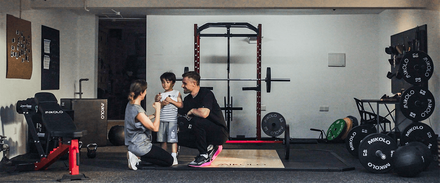
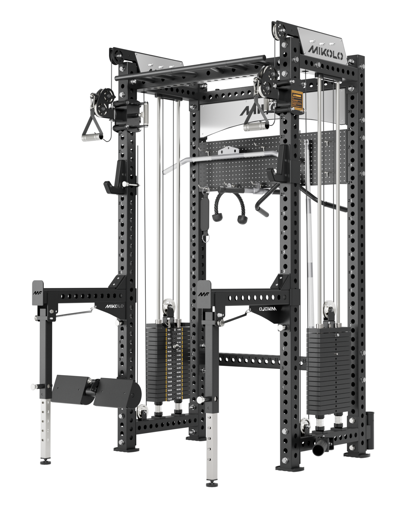
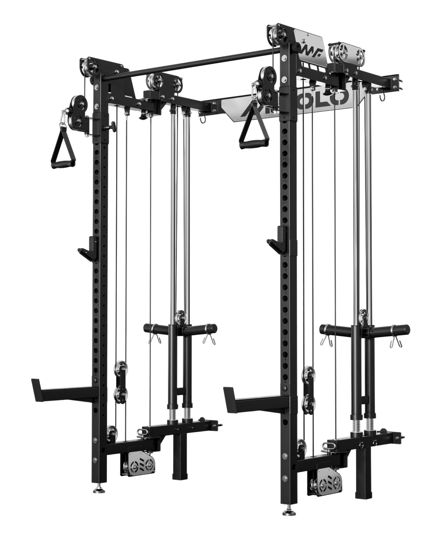
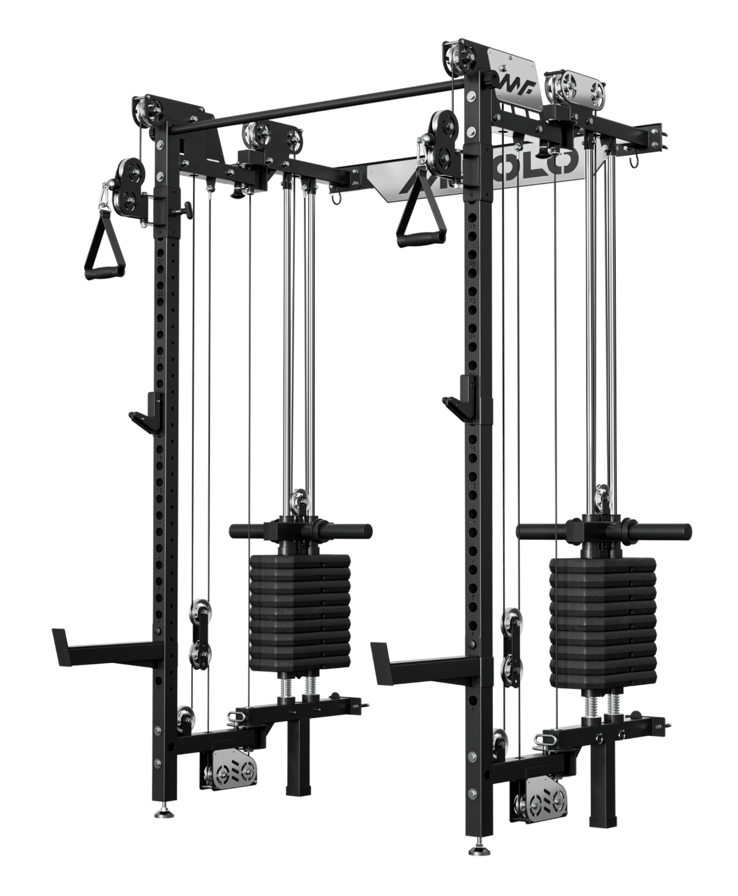
What Is a Squat Rack: How to Choose the Right One for Your Home Gym
If you're setting up a home gym, choosing the right squat rack can feel overwhelming. There are squat stands, half racks, power racks, and folding racks—and they all seem similar at first.But the...
What Do Preacher Curls Work: Complete Guide to Target Muscles and Benefits
Preacher curls are a go-to move for building bigger, stronger arms. Whether you’re using a preacher curl bench, dumbbells, or a cable machine setup, this exercise is designed to isolate your biceps...
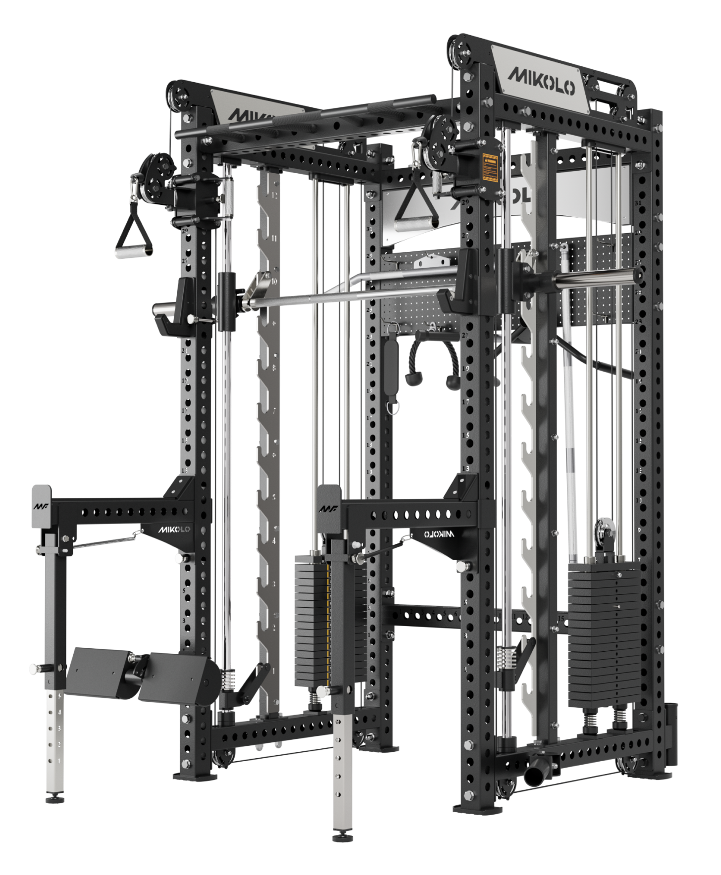
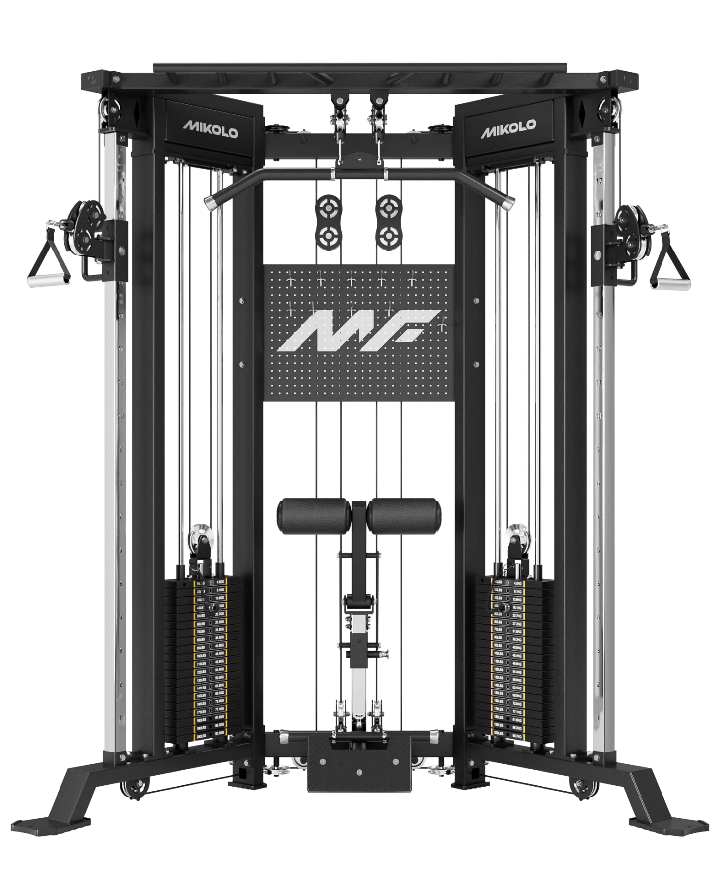
Smith Machine Guide: Benefits, Exercises & Best Options for Home Gym
The Smith machine is a staple in many gyms—a barbell fixed on steel rails, moving only up and down in a straight path. Unlike free weights, it guides your motion and comes with built-in safety hook...